
Age over 40
The risk increases with age. After 40 a check-up including eye pressure measurement and optic nerve assessment is advisable.
Ophthalmic surgeon · Catania, Sicily
Early diagnosis, laser and surgery for glaucoma with Dr Marco Di Maita. Consultations in English.
Centro Medico Polispecialistico Di Maita
Viale Alcide De Gasperi 151, Catania
01 Glaucoma in brief
Glaucoma is a disease of the optic nerve, the structure that carries visual information from the eye to the brain. In its early stages it often causes no symptoms: central vision stays good for a long time, while the visual field can slowly narrow at the edges.
The damage affects the fibres of the optic nerve. Raised eye pressure is the main risk factor that can be treated, but glaucoma can also occur with normal pressure.
Many people notice nothing for years. This is why glaucoma is often found during a routine check-up, before the person notices any change.
Vision that has been lost does not come back. Treatment aims to slow down or stop progression: finding glaucoma early allows treatment to start while there is more vision to protect.
02 Risk factors
Some conditions make glaucoma more likely. If one or more of these apply to you, it is worth discussing them with an ophthalmologist and planning regular check-ups.
The risk increases with age. After 40 a check-up including eye pressure measurement and optic nerve assessment is advisable.
Having a parent, brother or sister with glaucoma increases the risk: a good reason to start check-ups earlier.
It is the main risk factor that can be treated.
High short-sightedness calls for particular attention to the optic nerve.
People with diabetes already have regular eye checks: it is an opportunity to assess the risk of glaucoma too.
Steroid eye drops, sprays, creams or tablets, if used for a long time, can raise eye pressure in some people.
Having one or more risk factors does not mean you have glaucoma: it means regular check-ups are advisable.
03 Glaucoma check-up
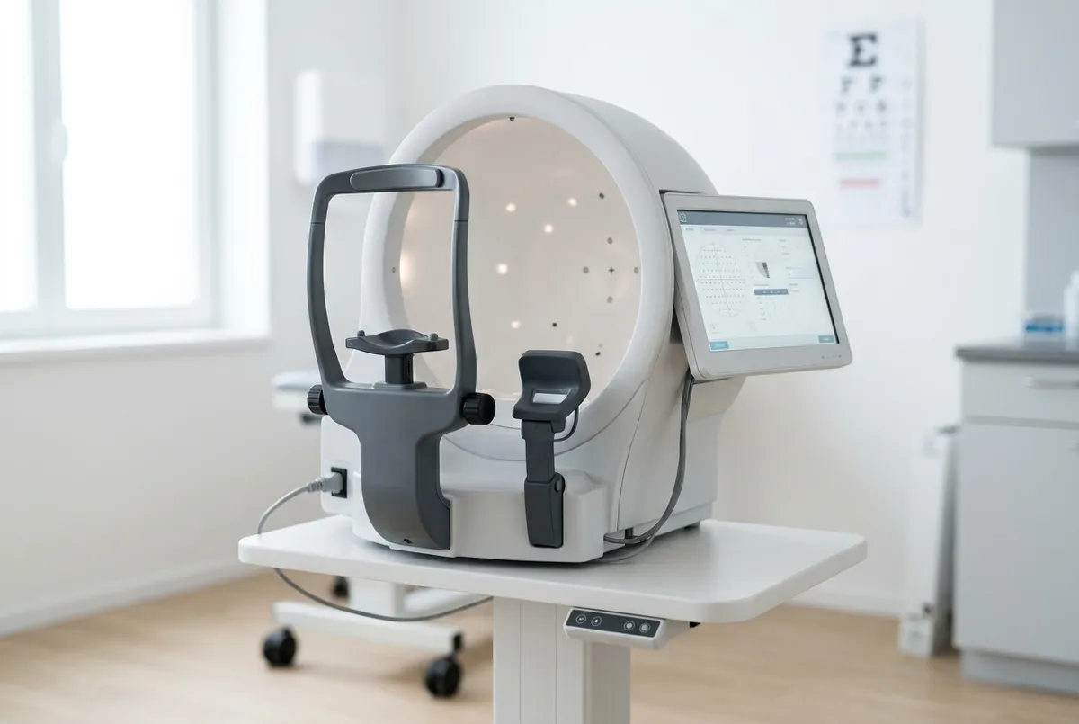
The check-up combines several tests, because no single test is enough to diagnose glaucoma or to monitor it over time. All the tests listed are carried out in the clinic.
Measures how well you see at distance and near, with the best correction from glasses or lenses.
Measures the pressure inside the eye. It is a quick and painless test.
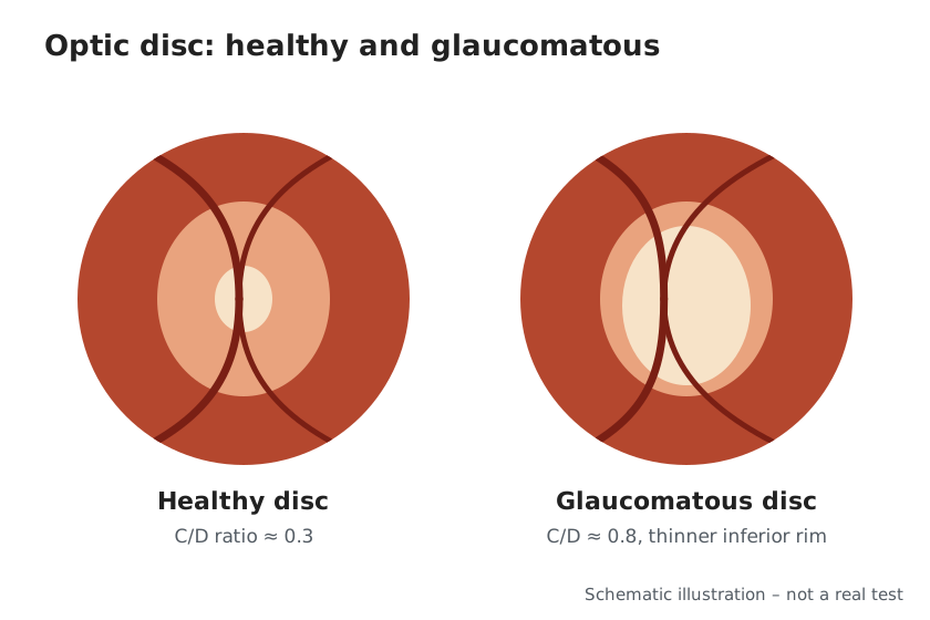
The ophthalmologist looks directly at the head of the optic nerve to assess its shape and colour.
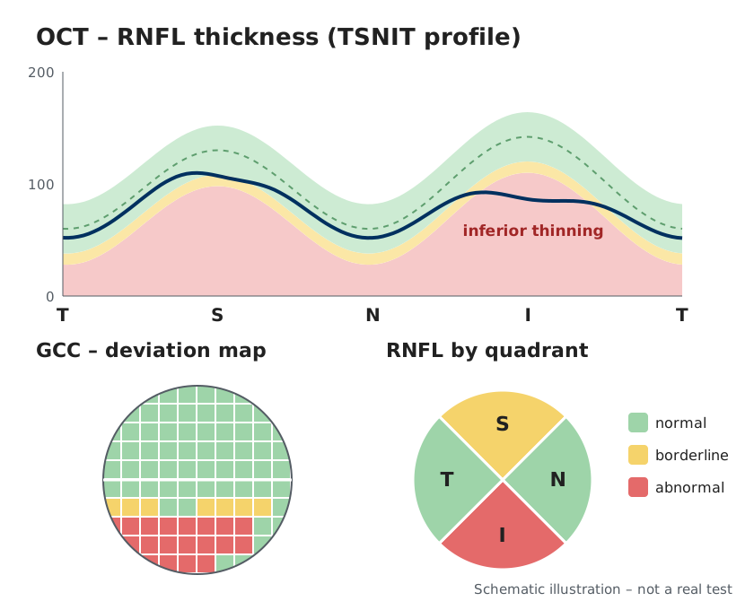
Measures the thickness of the nerve fibres and optic nerve and the ganglion cell layer of the retina. Ganglion cell analysis is especially useful for detecting early damage, while the visual field is still normal (pre-perimetric stage). It also allows results to be compared over time.
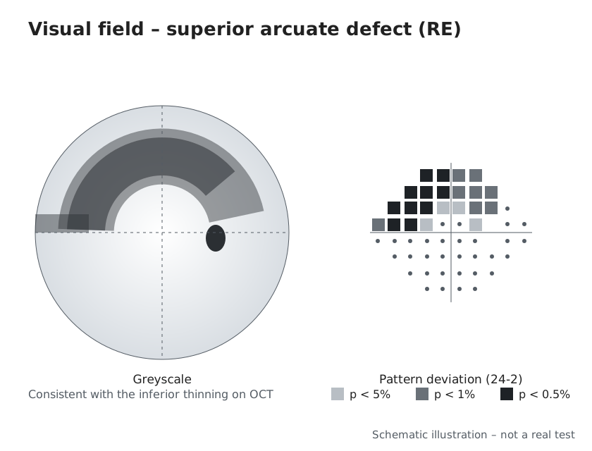
Assesses visual function across the different areas of the visual field, including the periphery, where glaucoma usually begins.
Measures the thickness of the cornea, which helps to interpret eye pressure correctly.
Examines the drainage angle of the eye, where the aqueous humour flows out, to distinguish between the different types of glaucoma.
These images are schematic illustrations for information only, not patient tests. They show a consistent example: thinning of the fibres in the lower part of the optic nerve corresponds to a defect in the upper part of the visual field.
 Tap to enlarge
 Tap to enlarge
 Tap to enlarge
04 Non-surgical treatments
In most cases glaucoma treatment starts without surgery. The aim is to bring eye pressure down to a target value set for each person.
These drops reduce the production of aqueous humour or help it drain. They work when used every day, regularly.
A low-energy laser acts on the trabecular meshwork, the structure through which the aqueous humour drains, to help it flow out. It is performed in the clinic, in a few minutes, with anaesthetic eye drops.
The LiGHT trial (Laser in Glaucoma and Ocular Hypertension). A randomised clinical trial carried out in the United Kingdom in patients with open-angle glaucoma or ocular hypertension compared SLT laser as first treatment with eye drops. In the SLT group about 3 in 4 patients did not need eye drops at 3 years and about 7 in 10 at 6 years, with a favourable safety profile. The results of a study are not a guarantee for the individual patient.
Selective laser trabeculoplasty versus eye drops for first-line treatment of ocular hypertension and glaucoma (LiGHT): a multicentre randomised controlled trial. Lancet. 2019;393(10180):1505-1516. doi: 10.1016/S0140-6736(18)32213-X
Laser in Glaucoma and Ocular Hypertension (LiGHT) Trial: Six-Year Results of Primary Selective Laser Trabeculoplasty versus Eye Drops for the Treatment of Glaucoma and Ocular Hypertension. Ophthalmology. 2023;130(2):139-151. doi: 10.1016/j.ophtha.2022.09.009
05 Glaucoma surgery
When eye drops or SLT laser are not enough to control eye pressure, or are not tolerated, glaucoma surgery is considered: trabeculectomy, microshunt, glaucoma drainage devices or, in mild to moderate glaucoma, minimally invasive procedures (MIGS), also together with cataract surgery.
What the choice of technique depends on
The pathway is not the same for everyone: in some cases surgery is the first step, in others eye drops or laser are enough.
01 Traditional surgery
The “classic” filtering surgery for glaucoma: an established technique that can achieve very low pressures.
It is a filtering operation: it creates a new drainage route for the aqueous humour under the conjunctiva, where a small collection of fluid called a filtering bleb forms. An antimetabolite, a medicine that reduces scarring, is often used.
When pressure is not controlled by eye drops or laser, or these are not tolerated. It is an established technique that can achieve very low pressures: for this reason it is considered when the target pressure is particularly low.
The operation is performed in the operating theatre. It requires closer post-operative follow-up, especially in the first weeks, when the bleb may need adjusting and the eye drop regime must be followed carefully.
02 Less invasive surgery
A tiny drain, with no tissue removed: less invasive glaucoma surgery, in selected cases.
A tiny drain carries the aqueous humour into a bleb under the conjunctiva, without removing tissue as in trabeculectomy.
When pressure is not controlled by eye drops or laser, or these are not tolerated, and a less invasive technique is considered appropriate for the target pressure to be reached.
It is a less invasive technique, with generally shorter operating times and a more favourable safety profile. In some studies, however, the pressure reduction is smaller or less long-lasting than with trabeculectomy. Regular check-ups are also needed after a microshunt.
Dr Di Maita is a co-author of a multicentre study on risk factors for failure after microshunt implantation (American Journal of Ophthalmology, 2024).
Risk Factors for Failure in Glaucoma Patients Undergoing Microshunt Implantation. Am J Ophthalmol. 2024;259:117-130. doi: 10.1016/j.ajo.2023.11.011
03 Tube and plate drainage devices
Devices with a tube and plate, considered mainly in more complex glaucoma or in eyes that have already had surgery.
A thin tube placed in the eye connects the anterior chamber to a plate positioned under the conjunctiva, towards which the aqueous humour drains.
It is typically considered in more complex glaucoma, in eyes with previous unsuccessful surgery, or when other techniques are less likely to succeed.
After implantation there may be a temporary phase of higher pressure before it stabilises. During this period eye drops and close check-ups may be needed.
04 Summary
| Feature | Trabeculectomy | Microshunt | Drainage device (tube) |
|---|---|---|---|
| Invasiveness | Higher: a small piece of tissue is removed and a filtering bleb is created | Lower: a tiny drain, with no tissue removed | Higher: implantation of a tube connected to a plate |
| Type of glaucoma in which it is considered | Glaucoma not controlled by eye drops or laser, including when a very low pressure is needed | Glaucoma not controlled by eye drops or laser, in selected cases | More complex glaucoma, eyes with previous unsuccessful surgery or where other techniques are less likely to succeed |
| Follow-up | Closer check-ups in the first weeks, then periodic | Regular check-ups in the first weeks, then periodic | Close check-ups, also because of the possible temporary phase of higher pressure |
| Main risks | Pressure too low, infection, bleb fibrosis, progression of cataract | Bleb fibrosis, pressure too low or insufficient reduction, further treatment | Pressure too low or too high, double vision, corneal problems, tube exposure |
05 Mild to moderate glaucoma
Minimally invasive procedures (MIGS) and SLT laser are used mainly in mild to moderate glaucoma, sometimes together with cataract surgery.
Procedures that improve drainage of the aqueous humour through very small incisions, with reduced surgical trauma and usually a quicker recovery.
Limitations: the pressure reduction is generally smaller than with filtering surgery; like any operation they carry risks, which are discussed during the consultation.
An outpatient treatment lasting a few minutes, with anaesthetic eye drops, that helps the aqueous humour drain. It can be the first treatment, as shown by the LiGHT trial, or be combined with eye drops.
Limitations: the effect may wear off over time; in some cases the treatment can be repeated.
In selected cases cataract surgery can be combined with a glaucoma procedure, for example MIGS, in the same session.
Limitations: whether this is appropriate depends on the type and stage of glaucoma and is assessed after examination and tests.
Would you like to know whether glaucoma surgery is appropriate in your case?
The surgical assessment includes a consultation and the check-up tests.
06 The doctor
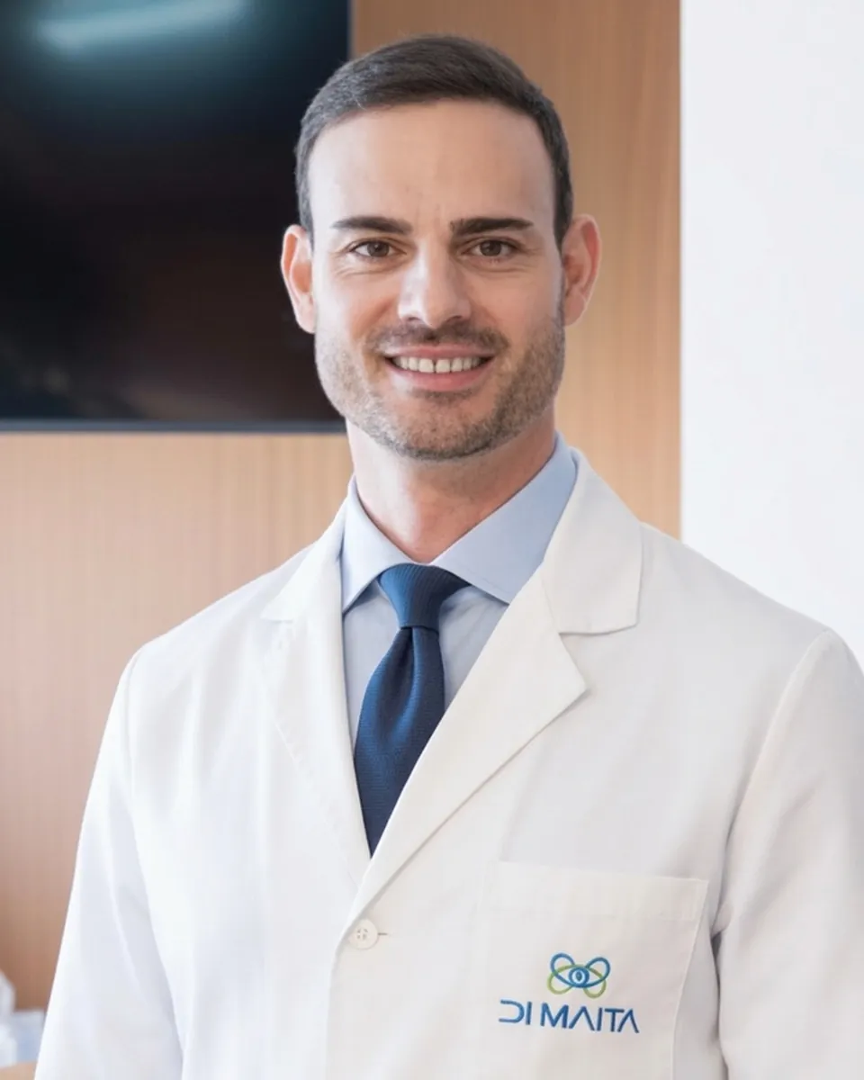
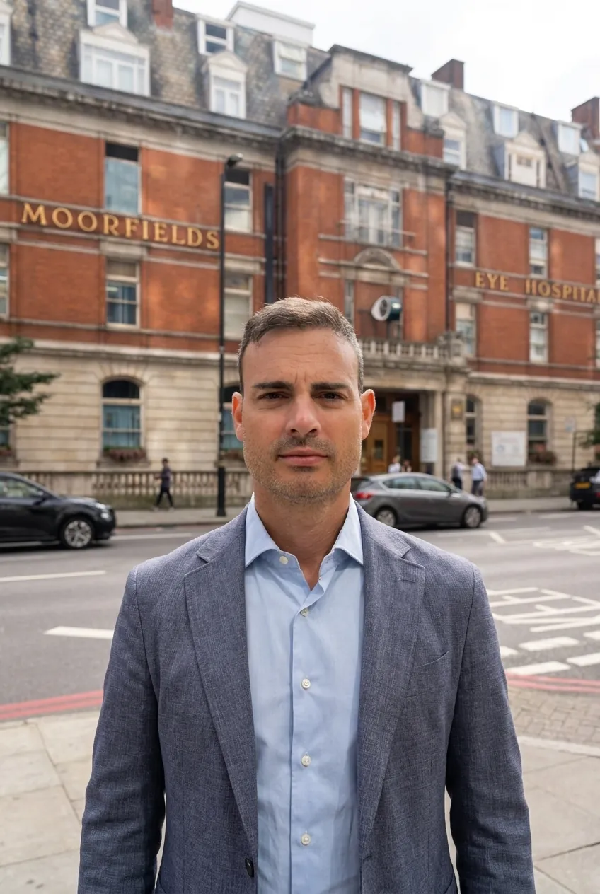
Dr Marco Di Maita is an ophthalmic surgeon. He graduated in Medicine and Surgery from the University of Catania and specialised in Ophthalmology at the University of Genoa in 2019, with honours. In the same year he became a Fellow of the European Board of Ophthalmology (FEBO) in Paris.
From 2021 to 2022 he was head of the Glaucoma Centre at Advalia Vision in Milan. Between 2023 and 2024 he completed a clinical and surgical glaucoma fellowship at Moorfields Eye Hospital in London. He is a member of the European Glaucoma Society.
He has performed thousands of cataract and refractive operations and numerous glaucoma treatments, including minimally invasive surgery. He is a co-author of papers in international journals (British Journal of Ophthalmology, European Journal of Ophthalmology, American Journal of Ophthalmology) and takes part in national and international conferences.
He speaks English and French. He sees patients in Catania at Centro Medico Polispecialistico Di Maita, where he performs surgery privately. He also operates both under the Italian National Health Service (SSN) and privately at Policlinico Morgagni in Catania.
07 Other areas
In addition to glaucoma, Dr Di Maita performs cataract surgery and refractive surgery.
A cataract is the clouding of the crystalline lens, the eye's natural lens. The operation replaces the clouded lens with an intraocular lens, chosen according to the measurements of the eye and the person's visual needs. Like any operation, it carries risks, which are explained during the consultation. In people with glaucoma, cataract surgery can, in selected cases, be combined with a procedure for eye pressure.
Refractive surgery corrects vision problems such as short-sightedness, long-sightedness and astigmatism, with laser or with intraocular lenses. Before it is offered, specific tests check whether the eye is suitable: not everyone is, and the result can vary from case to case.
08 Frequently asked questions
Short answers to the most common questions. Each situation still needs to be assessed during a consultation.
The information on this website is for general information only and does not replace a specialist consultation.
No: damage that has already occurred to the optic nerve cannot be reversed. The aim of treatment (eye drops, laser or surgery) is to slow down or stop progression, to preserve the vision you have. This is why early diagnosis matters so much.
After the age of 40 an eye examination with eye pressure measurement and optic nerve assessment is advisable, repeated as often as the ophthalmologist recommends based on your risk factors. People already diagnosed with glaucoma follow a personalised follow-up schedule.
Usually not. It is performed in the clinic with anaesthetic eye drops and takes a few minutes. You may feel mild discomfort or temporary glare; afterwards you go home.
Both create a drainage route for the aqueous humour under the conjunctiva. Trabeculectomy removes a small piece of tissue, can achieve very low pressures and requires closer follow-up. A microshunt uses a tiny drain without removing tissue and is less invasive, but in some studies the pressure reduction is smaller or less long-lasting. The choice is made case by case.
It is considered mainly in more complex glaucoma, in eyes with previous unsuccessful surgery, or when other techniques are less likely to succeed. The decision is made after examination and tests.
Glaucoma surgery is usually performed under local anaesthesia as day surgery, with no overnight stay. The type of anaesthesia is decided according to the person and the procedure.
This cannot be known in advance. After surgery some people reduce or stop their pressure-lowering drops, while others need to keep using one or more. In the weeks after surgery post-operative drops are used in any case.
In selected cases, yes: cataract surgery can be combined with a glaucoma procedure, for example a minimally invasive one (MIGS). Whether this is appropriate depends on the type and stage of glaucoma and is assessed after examination and tests.
A private first eye examination costs from €130. Any additional tests depend on the clinical picture; for detailed information please contact the front desk.
Yes: Dr Di Maita operates both under the Italian National Health Service (SSN) and privately at Policlinico Morgagni in Catania. At Centro Medico Polispecialistico Di Maita he performs surgery privately. For access arrangements and waiting times please contact the front desk.
09 Location and opening hours
Dr Di Maita sees patients at DI MAITA – Centro Medico Polispecialistico. To book you can use MioDottore, call, message on WhatsApp or write using the form.
| Monday | 14:00 – 19:00 |
|---|---|
| Tuesday | 9:00 – 19:00 |
| Wednesday | 9:00 – 14:00 |
| Thursday | 9:00 – 19:00 |
| Friday | 9:00 – 14:00 |
| Saturday and Sunday | Closed |
10 Write to us
The front desk will contact you on the days the clinic is open. You can write in English. For urgent requests please call +39 351 429 5888 or message on WhatsApp.
To protect your privacy, please do not include medical reports or detailed clinical information in your message: you will discuss them directly with the doctor.